Assistant Professor, Mercer University School of Medicine
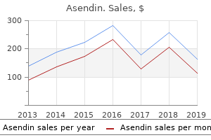
Safe 50 mg asendin
Preoperative Planning the injured hip should be evaluated underneath anesthesia utilizing fluoroscopy anxiety feeling asendin 50 mg for sale. Approach Extra-articular fractures which might be secure after discount must be immobilized in a spica solid anxiety vs fear 50 mg asendin mastercard. Many neck fractures could be reduced closed and fixed percutaneously from laterally depression definition gdp 50 mg asendin free shipping. If the fracture could be anatomically reduced lithium depression definition purchase cheap asendin on line, the surgeon ought to proceed with percutaneous fixation; if not, open reduction must be undertaken. After reduction, pins are drilled from the lateral femoral cortex retrograde throughout the fracture. C D It is customary to use clean pins for physeal separations or neck fractures in very young children. A lateral view, often by frogging the hip, is critical to affirm pin placement. Because there could also be a tense hemarthrosis that tamponades flow in the retinacular vessels of the neck, it might be sensible to aspirate the joint capsule to evacuate or decompress the hip joint. The goal is anatomic reduction to preserve perfusion to the capital femoral epiphysis, optimize bony apposition for healing, and forestall deformity, particularly varus and exterior rotation. The vastus lateralis is incised longitudinally, and the muscular tissues overlying the anterior hip capsule are elevated anteriorly. The incision is curved posteriorly and then extends distally in the posterior third of the vastus fascia. The angle the guidewire makes with the femoral shaft is dictated by the fixation device to be used. The fascia of vastus lateralis is "hockey sticked" and vastus muscle is retracted anteriorly, exposing the lateral femoral cortex. After fracture reduction, a guidewire is inserted from the lateral femoral cortex up the femoral neck. The angle the wire makes with the lateral cortex should match the angle of the fixation gadget (usually a hundred thirty five degrees). Reaming is accomplished over the guidewire to accommodate the lag screw and the barrel of the aspect plate. The plate is secured to the femur with cortical screws and the compression screw locks the lag screw within the facet plate. The surgeon should consider decompressing the hemarthrosis to reduce the impact of tamponade of the vessels. Parents are warned in advance of the likelihood and implications of avascular necrosis. Perfect discount and bony apposition provide the best alternative for fracture healing. Decompression and stable inner fixation of femoral neck fractures in children can affect the finish result. In children who maintain multiple traumatic accidents, the character and severity of each harm have to be considered to optimize treatment. The proximal ossification heart is seen by 6 months and the distal femoral ossification center appears at 7 months. The profunda femoris artery provides rise to four perforating arteries, which enter the femur posteromedially. During fracture therapeutic, however, nearly all of the blood is provided by the periosteal circulation. The diploma of trauma required to trigger harm will increase exponentially because the character of the bone modifications and steadily turns into stronger and bigger from infancy to adolescence. Low-energy injuries resulting in fractures may level to a pathologic nature of the condition, except in toddlers, in whom low-energy femur fractures are frequent. The place of the fracture fragments after the injury depends on the level of the fracture and displays the gentle tissue and muscle forces performing on the femur. In the setting of an isolated femur fracture, the thigh seems swollen with minor bruises and abrasions.
Asendin 50mg otc
Concurrent fractures depression quest steam cheap 50 mg asendin free shipping, mostly involving the distal radius depression symptoms not sad buy generic asendin, scaphoid depression definition and meaning 50mg asendin free shipping, and proximal humerus depression definition emedicine purchase generic asendin canada, occur in 1% of circumstances. Associated neurovascular accidents can happen, with preoperative nerve injury current in 8% of circumstances and vascular insufficiency present in 1% to 2% of cases. During a fall with the elbow in full extension, the olecranon in its fossa acts as a fulcrum. The capsule, because it inserts distal to the olecranon fossa and proximal to the physis, transmits an extension force to this region, leading to failure and fracture. With the elbow in full extension and the elbow turning into tightly interlocked, bending forces are concentrated within the distal humeral region. Increased ligamentous laxity, resulting in hyperextension of the elbow, could also be a contributing factor to this injury sample. The majority of supracondylar fractures of the humerus (other than extension kind I fractures) are unstable; due to this fact, stabilization within the type of forged immobilization or ideally operative fixation is normally needed. Forearm supination normally aids within the reduction of these posterolaterally displaced fractures. Lateral displacement of the distal fragment locations the median nerve and brachial artery in danger. The ulnar nerve courses via the cubital tunnel posterior to the medial epicondyle. It is at explicit threat with flexiontype fractures or when a medial pin is positioned for fracture fixation. Therefore, the elbow should be relatively extended if a medial pin is placed for fracture fixation. The physical examination could reveal swelling, tenderness, ecchymosis, and deformity. The pucker signal, which occurs on account of the proximal fracture fragment spike penetrating via the brachialis and anterior fascia into the subcutaneous tissue, may be present. Physical examinations to carry out embody: Assessing for potential related injury to the ulnar nerve. Finger, wrist, and thumb extension (extensor digitorum communis, extensor indicis proprius, extensor carpi radialis longus and brevis, extensor carpi ulnaris, extensor pollicis longus) is examined. The proximal metaphyseal spike penetrates laterally with posteromedially displaced fractures and locations the radial nerve at risk. With posterolaterally displaced fractures, the spike penetrates medially and places the median nerve and brachial artery in danger. Index distal interphalangeal flexion (flexor digitorum profundus index) and thumb interphalangeal flexion (flexor pollicis longus) are examined. Nonoperative management consists of immobilization of the elbow in no more than ninety levels of flexion in a splint or solid. Historically, some supracondylar fractures of the humerus were managed with traction (overhead versus side). With the relative safety of percutaneous pinning strategies, however, using traction has been limited. Biomechanical studies have revealed comparable stability within the lateral-entry and crossed-pin strategies. The presence of a posterior fat-pad sign suggests an intra-articular effusion and could be related to an occult supracondylar fracture of the humerus. On a lateral view of the elbow, the anterior humeral line should intersect the capitellum. An advantage of the lateral-entry pin method is the significantly decrease risk of iatrogenic nerve harm. The crossed-pin technique could also be indicated if persistent instability is famous intraoperatively after placement of three lateral-entry pins. Indications for open reduction of supracondylar fractures of the humerus are limited but embrace open accidents, fractures irreducible by closed means, and fractures associated with persistent vascular compromise even after sufficient closed discount. Complete preoperative neurologic and vascular examination is carried out and documented. The contralateral arm must be examined, and the carrying angle of the contralateral arm ought to be famous. Recent retrospective studies recommend that a delay in therapy of the majority of supracondylar fractures is appropriate.
Purchase discount asendin line
Orientation of the transfer tunnel between the medial and anterior pores and skin incisions (red arrow) bipolar depression children buy asendin pills in toronto. A clamp is positioned into the tunnel from distal medial to proximal anterior and used to information the rectus femoris muscle�tendon unit to its web site of switch insertion (circle) mood disorder symptoms in children generic 50 mg asendin overnight delivery. The rectus femoris tendon is delivered into the medial incision (solid circle) anxiety 1st trimester asendin 50 mg for sale, where it is going to be transferred to the distal portion of the semitendinosus muscle tendon (dashed circle) anxiety xanax forums discount asendin 50mg with amex. The transfer is tensioned in order that the muscle belly of the rectus femoris muscle is barely tighter to palpation than the muscle bellies of the remaining three muscular tissues of the quadriceps muscle group, when the knee is held in full extension. The rectus femoris muscle is a biarticular muscle and ought to be transferred to another biarticular muscle (such because the semitendinosus). The line of pull of the transferred muscle�tendon unit must be as straight as possible. The lengthy anterior thigh incision and intermuscular proximal release of the rectus femoris muscle must be performed. The transfer tunnel for the rectus femoris muscle should be on the degree of the subcutaneous fats, superficial to the quadriceps fascia. The muscle switch must be tensioned so the muscle belly is at a slight stretch, to optimize the length�tension relationship of the transferred muscle. The rectus femoris muscle switch must be tensioned so the muscle is barely tighter than the opposite parts of the quadriceps muscle group. If full knee extension has been achieved after lengthening of the medial hamstring muscular tissues and switch of the rectus femoris muscle, then the knee is protected in a knee immobilizer after surgery. The knee immobilizer is worn full time and the child is saved non-weight bearing for 2 weeks. This should be corrected by acceptable gait coaching early within the rehabilitation part. Improved dynamic alignment on the knee during the swing phase of the gait cycle should end in improved gait effectivity and clearance of the swing limb. Improvements in swing-phase knee kinematics after rectus femoris muscle switch have been documented at 1 year after surgical procedure and have been proven to be maintained at 5 and 10 years of follow-up. Proper rehabilitation under the course of an skilled bodily therapist is efficient in managing this drawback. The principal beauty complication after switch of the rectus femoris muscle is an unpleasant scar which will develop on the incision web site on the anterior facet of the thigh. This is a consequence of the popular incision crossing the pores and skin lines of Langer. Scar formation is minimized by proper incision wound management (pressure applied by massage) through the postoperative rehabilitation section. Prediction of end result after rectus femoris surgical procedure in cerebral palsy: the role of cocontraction of the rectus femoris and vastus lateralis. Diminished knee flexion after hamstring surgery in cerebral palsy: prevalence and severity. Force and second producing capacity of the lower extremity muscular tissues earlier than and after tendon lengthening. Knee motion following multiple soft tissue releases in ambulatory sufferers with cerebral palsy. Treatment of stiff-knee gait in cerebral palsy; a comparability of distal rectus femoris transfer versus proximal rectus launch. Propulsive perform during gait in diplegic youngsters: analysis after surgery for gait improvement. Based on modeling studies, the hamstrings are a significant contribution to increasing the drive in spastic hip illness, which causes hip subluxation. They are also a part that retains the knees flexed and secondarily encourages flexion combined with spastic hip flexors, which causes the knee to fall into internal rotation and adduction, magnifying the affect of the concomitant spastic adductors. This posture of hip flexion and inner rotation and adduction, with the addition of high muscle pressure, tends to drive the hip posterosuperiorly out of the acetabulum. The primary period throughout which spastic hip illness occurs is 2 to 8 years of age, although some kids are nonetheless at risk by way of their adolescent development spurt and need to be monitored. The main physical examination discovering is the limitation of hip abduction with hips extended and knees extended.
Asendin 50 mg mastercard
Anterior cruciate ligament reconstruction in skeletally immature knees: an anatomical examine 9435 mood disorder purchase asendin without prescription. The conservative treatment of full tears of the anterior cruciate ligament in skeletally immature sufferers mood disorder definition 50mg asendin free shipping. Anterior cruciate ligament reconstruction autograft choice: bone-tendon-bone versus hamstring: does it really matter Clinical longitudinal standards for peak bipolar depression organizations generic asendin 50 mg with amex, weight mood disorder hcc buy asendin online, height velocity, weight velocity, and phases of puberty. Healing was noted within the medial femoral condyle in 3 of 10 patients; therapeutic elsewhere was famous in 10 of eleven patients. In late presentations by which an osteochondral flap or unfastened body is current, basic biomechanical symptoms together with locking, catching, buckling, and giving means could happen. With careful palpation through various amounts of knee flexion, some extent of maximal tenderness often may be situated over the anterior medial side of the knee. The tender area corresponds to the lesion, usually on the lateral facet of the distal medial femoral condyle. With steady lesions, knee effusion, crepitus, and extreme pain by way of a normal range of motion are rarely observed. The tibia is then internally rotated because the knee is prolonged from ninety degrees toward full extension. In a optimistic Wilson check, ache is elicited over the anterior aspect of the medial femoral condyle. The mechanical symptoms are extra pronounced in the unusual circumstance in which the kid or adolescent presents with an unstable lesion. In steady and unstable shows, both knees ought to be examined to determine whether the situation is bilateral. Ipsilateral quadriceps atrophy can also be noted if the affected person has been having pain for more than an prolonged time period. Some patients are believed to have a genetic, biochemical, or behavioral predisposition towards this situation. The targets of imaging are to characterize the lesion, determine the prognosis of nonoperative management, and monitor the healing of the lesion. Tunnel view of the knee is especially useful in lesions over the flexion floor of the medial femoral condyle. Plain radiographs usually characterize and localize the lesion and rule out different bony pathology of the knee area. Controversy exists regarding the ideal nonoperative management for these patients. Clinicians who adhere to treating the subchondral bone as the first supply of pathology favor a interval of immobilization. Those whose focus is on the articular cartilage as a source of pathology are inclined to favor sustaining mobilization. The choices for immobilization embrace casting, bracing, and standard knee immobilization. The brace could also be unlocked to work on range of movement for five minutes 5 instances per day. High-impact actions and activities that may involve shear stress to the knee ought to be restricted till the kid has been pain-free for several months and the radiographs present a healed lesion. The aim of nonoperative intervention is to promote therapeutic within the subchondral bone and potentially forestall chondral collapse, subsequent fracture, and crater formation. The targets of operative remedy are to promote therapeutic of subchondral bone, to maintain joint congruity, to repair rigidly unstable fragments, and to replace osteochondral defects with cells that may replace and grow cartilage. Optimal surgical treatment supplies a steady assemble of subchondral bone, calcified tidemark, and repair cartilage with viability and biomechanical properties equal to or similar to native hyaline cartilage. The leg can be placed in a leg holder on the operating desk with the knee joint previous the tip of the operating desk, thus permitting the knee to flex 90 levels and the decrease leg to grasp freely. The leg may be positioned supine on the working desk, with the hip flexed and the knee flexed ninety degrees.
Safe 50 mg asendin. Unemployed Depressed and Terrified.
Order 50mg asendin amex
Preoperative Planning A detailed evaluation of the scientific findings and all appropriate imaging studies is performed earlier than the procedure depression lies buy generic asendin 50 mg online. Shortening must be determined to be lower than 2 cm using a lateral radiograph major depression clinical definition asendin 50 mg on-line, although some recommend spica casting may be completed regardless of shortening anxiety effects cheap asendin 50mg with mastercard. In infants vegetative depression definition buy 50 mg asendin fast delivery, stable femoral shaft fractures could be treated in a Pavlik harness or a splint. In kids youthful than 6 years, closed reduction and casting is used within the overwhelming majority of circumstances. Positioning the kid is taken to the working room or sedation unit and positioned in the supine place on the desk. The injured extremity is casted first, after which the affected person is transferred to a spica desk. Because of recent stories of compartment syndrome of the leg after spica casting for pediatric femur fractures,8,9 many facilities (ours included) have been using less hip and knee flexion and not including the foot for the forged of the injured leg. To avoid vascular compromise, care should be taken not to flex the knee as soon as the padding is in place. The patient is transferred to a spica desk, the place the load of the legs is supported with manual traction. The remainder of the spica solid is placed whereas holding the fracture out to size. Care must be taken to avoid extreme traction, which increases the danger of compartment syndrome and pores and skin sloughing. Gore-Tex liners are used at some institutions to prevent diaper rash and superficial infections. Traditional spica casting with 90 degrees of hip flexion, 30 degrees of abduction, and 15 levels of external rotation. The foot stays uncovered with the cast stopping within the supramalleolar area, which is protected with extra padding. The pelvic band is utilized with a number of layers of stockinette folded on the stomach to prevent abdominal compression from the casting. Seven or eight layers of folded fiberglass are positioned within the inter-hip crease to lower the risk of the cast breaking, whereas a wide pelvic band is needed to immobilize the hip as nicely as attainable. Cylinder forged with 50 degrees of knee flexion and 45 degrees of hip flexion for walking spica forged. Walking spica casting position with 30 degrees of abduction and 15 levels of exterior rotation. Wide pelvic band and anterior reinforcement for additional help in a final strolling spica solid. Shortening of greater than 2 cm (controversial) Massive swelling of the thigh Associated harm that precludes solid remedy Walking spica Effective for low-energy isolated femur fractures Toddlers typically pull-to-stand and start strolling in 2 to three weeks. We counsel the household, immediately after discount in casting, that wedging of the solid may be necessary at about 10 to 14 days after harm. This frequently avoids pointless trips again to the working room in the postoperative period for loss of reduction. Prior to callus formation, if shortening of more than 2 cm occurs, one of three options could also be required: cast change, traction, or external fixation. Shortening of more than 2 cm once callus has fashioned may be handled with osteoclasis and lengthening techniques at a tempo of 1 mm per day. Illgen and colleagues6 found that commonplace spica casting was profitable (without forged change or wedging) about 86% of the time. Immediate spica casting in the emergency division under conscious sedation and discharge has been proven to have related charges of complication and re-reduction as "early" spica casting. Epidemiology of femoral fractures in children in the West Midlands region of England 1991 to 2001. Spica solid software within the emergency room for choose pediatric femur fractures. Fractures of the femoral shaft in kids: incidence, mechanisms, and sociodemographic danger factors. Compartment syndrome of the leg after therapy of a femoral fracture with an early sitting spica forged: a report of two instances. Volkmann contracture and compartment syndromes after femur fractures in kids treated with 90/90 spica casts.
Buy asendin 50mg low price
Autograft typically is taken from the iliac crest (ie anxiety rash purchase asendin 50mg on line, tricortical iliac crest graft) Allograft combines tricortical iliac crest with croutons for medullary packing mood disorder types purchase asendin with american express. Pre-incision fluoroscopy images should be obtained of the hip and ankle to ensure that the operative bed can accommodate access to these areas geriatric depression definition quality asendin 50 mg. The hip and ankle are wanted for intraoperative evaluation of the mechanical axis depression symptoms test online proven asendin 50mg. Approaches for the lateral closing wedge are from the anterolateral side of the tibia, simply anterior to the fibula. A lateral submit is used through the arthroscopy portion of the procedure and could be lowered in the course of the open osteotomy portion. If the osteotomy is being carried out along with a cartilage restorative process (eg, autologous chondrocyte implantation), the osteotomy is performed first and then the restorative cartilage procedures are carried out, to decrease any trauma to the newly implanted periosteal overlaying or injected cartilage cells. On entering the lateral compartment, an sudden cartilage lesion was discovered on the lateral femoral condyle. Offloading the mechanical axis into the lateral compartment that already is degenerated is a contraindication to the process. The tibial tubercle, posteromedial tibia, and joint line are clearly identified with a skin marker. The superior border of the gracilis hamstring tendon is palpated, and the sartorius fascia is opened along the superior border of the gracilis tendon. Medially, the pes bursa is released from the medial tibial tubercle in an inverted L style. The pes bursa is rigorously elevated distally, taking great care to develop the aircraft between the bursa and the underlying medial collateral ligament. Anteriorly, the patellar tendon is recognized, and a aircraft posterior to the tendon is recognized. Occasionally, the most superior fibers of the patellar tendon attachment to the tibial tubercle must be elevated to keep away from inadvertent creation of the osteotomy through the patellar tendon. The Cobb elevator is then used to dissect the muscular tissues and tissues from the posterior tibia alongside the road of the osteotomy. Care must be taken to stay immediately on the posterior tibial bone to keep away from neurovascular harm. After enough posterior dissection, it ought to be possible to move a finger bluntly across the posterior tibia. For additional protection of the posterior neurovascular buildings, a laparotomy sponge is positioned across the again of the knee. Overall orientation of incision by way of subcutaneous fats, all the method down to sartorius fascia. The sartorius fascia is opened just superior to the gracilis tendon, and the pes bursa is elevated off in an L-type style. Before the osteotomy is performed, an intraoperative mechanical axis view should be obtained, using both the Bovie twine or the alignment rod found in the osteotomy set. The angle of the information pin assembly is changed in order that the information pins are just superior to the tibial tubercle. Two pins are drilled from medial to lateral alongside the osteotomy line to intersect the preliminary information pin 1 cm from the lateral cortex. Fluoroscopic image verifying the two information pins placed from medial to lateral utilizing the osteotomy guide pin assembly. Note how on this view, which is parallel to the joint floor, the two pins are superimposed on each other, thus verifying that they, too, are parallel to the joint surface. White arrow, information pin meeting; black arrow, osteotomy guide pins; black arrowhead, initial guide pin. A guide pin is positioned from medial to lateral throughout the proximal tibia, 1 cm distal to the joint, and parallel to the joint floor. Not solely can the osteotomy information pin meeting decide the angle of the reduce in the coronal aircraft, but it also has the power to rotate within the sagittal aircraft to reproduce the anterior-to-posterior tibial plateau slope accurately. The angle of the guide pin assembly within the coronal plane is about in order that the information pins will enter the proximal tibia above the tibial tubercle. When acceptable, two additional information pins are drilled from medial to lateral along the orientation of the osteotomy reduce. The parallel information sleeve, information pin meeting, and preliminary guidewire parallel to the joint line at the second are eliminated. Either with or with out the cutting guide, an oscillating noticed is used to make the osteotomy reduce.
Purchase 50 mg asendin with visa
The spinous processes of the cranial and caudal foundations are exposed and marked with a metallic object similar to a Kocher clamp bipolar depression journal articles discount asendin express, and a lateral radiograph is then used to confirm the degrees mood disorder 5 year old buy 50 mg asendin with mastercard. They are composed of a minimum of two pair of anchors and usually span two or three vertebral levels depression symptoms and definition buy generic asendin 50mg on line. The foundations include the vertebral segments at both ends of the constructs depression with symptoms of psychosis order asendin from india, which are internally fastened with anchors. Limited fusions of the muse ranges typically are carried out utilizing native bone graft or allograft extenders to provide extra stability. The posterior components of the cranial and caudal foundations are exposed subperiosteally out to the extent of the transverse processes. Pedicle screws also may be used, normally with four screws spanning two vertebral levels. Pedicle screws could offer increased stability to the construct and are most popular for each foundations so long as the anatomy allows their secure placement. In basic, the thoracic pedicle start line is located at the intersection of the lateral border of the superior articular facet and the cranial aspect of the transverse course of. Lumbar pedicle screws start on the junction of the pars interarticularis, the midpoint of the transverse course of, and the base of the superior articular process. Fluoroscopy and neural monitoring are useful in aiding pedicle screw placement, particularly in sufferers with deformity. Adding the Rods Placing the Anchors Once exposure has been obtained, the anchors are positioned. Contralateral supralaminar hooks may be staggered over two levels if canal stenosis is a concern. Next, upgoing hooks are placed, usually underneath the side articulations of the same vertebra however typically in a staggered association. Foundations utilizing supralaminar hooks usually encompass three vertebral ranges with supralaminar hooks placed on either aspect of the cranial two vertebra and the side hooks placed on the most caudal one. For extra stability, further facet hooks can be utilized to prolong the inspiration to three ranges. The twin rod technique employs two rods, every made up of a cranial basis rod and a caudal basis rod joined by a connector, for a complete of 4 rods for the complete construct. Either of two types of connectors could also be used: a tandem connector, which houses the cranial and caudal rods inside an oblong field so the ends meet finish to finish, or side-to-side connectors, which allow the rods to overlap. If any contouring is important within the area where the cranial and caudal rods meet, closed twin connectors must be used, with an overlap of 2 to four inches to allow future lengthening. Bilateral rods are ready for each foundation by measuring the length of the spine in the corrected position and carefully contouring the rod. Subfascial placement involves a much deeper dissection, both initially and with every lengthening, nevertheless, and may enhance the chance of premature fusion. Subcutaneous placement could additionally be associated with the next incidence of skin issues and wound an infection. A single pores and skin incision could also be used with subperiosteal publicity of the cranial and caudal basis websites. The lateral view exhibits the straight tandem connector positioned within the thoracolumbar region. The trajectory of the pedicle screws can also be seen and varies between sufferers. Close-up of the cranial foundation exhibits 4 pedicle screws spanning two ranges in the thoracic area. Close-up of the caudal foundation exhibits four pedicle screws spanning two levels within the lumbar spine. Growing rod technique for the therapy of progressive early onset scoliosis in fusionless surgical procedure for backbone deformity. These transverse connectors enhance the soundness of the assemble, particularly when hooks have been used. The cranial anchors and transverse connector are tightened first, adopted by the caudal anchors and transverse connectors. Cranial and caudal set screws are positioned on the side of the tandem connector that correlates to the most distinguished facet. Next, the rod construct on the convex side of the curve is created equally and tightened.